
When you think of “integumentary” what do you think of? Well if you are anything like me you think, “Dang, that’s a long confusing word…must be Science related!!” and if you thought that, you would be correct! The Integumentary system is definitely a science topic! The short answer to “what is the integumentary system?” would be that its skin. But the skin is the biggest organ in your body! (Yes, it IS an organ!) And so being a HUGE organ, there are many parts to it. Look at your hair, now back at this. Look at your nails. Now back at this, I’m orange now, now look at the color of your skin, now back at me, I’m black again! All of these features you just looked at are all part of the integumentary system.
So let’s begin with the layers of the skin that we all have!
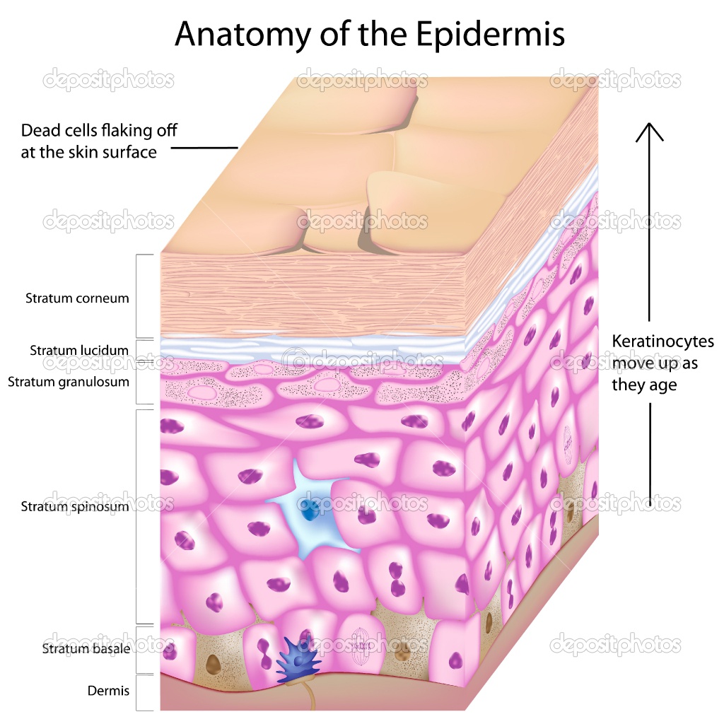
Epidermis: The epidermis is the outermost layer of the skin. The epidermis is comprised of Keratinocytes, Melanocytes, Langerhans, and Merkel cells! Human skin is continually being renewed, in contrast with that of reptiles that molt. The desquamation of cells on the skin’s surface should naturally be compensated for by renewal of the epidermis, a process undertaken by the Keratinocytes (85% of the cells in the epidermis). These possess two properties which successively come into action – the ability to actively divide and the ability to differentiate.
Horny Layer: the first layer of the Epidermis
Clear Layer: Second layer of the Epidermis (guess what! It’s clear!!!)
Granular Layer: third layer, skin cells begin to die here and rise until they reach the horny layer. This is also the thinnest layer of the epidermis!
Prickly Layer: 4th layer (second deepest layer)
Basal Layer: 5th layer, the deepest layer of the epidermis!
To get a better look…here is a picture!!
The next big layer is the dermis! The dermis is 10 to 40 times thicker than the epidermis. At the junction with the epidermis, its surface bristles with fibrous, vascular and nervous projections – the dermal papillae. The fibroblasts are the main cells in the dermis. They are essentially located in the dermal papillae close to the epidermis, and found only in very low numbers in the deep layers of the dermis known as the reticular dermis. They are specialized in producing two types of protein fibers, collagen and elastin fibers constituent of the extra-cellular matrix. Collagen fibres, 70% of the proteins in the dermis, give the dermis its resistance to strain and traction, while elastin supplies its elastic properties.
The reticular dermis accounts for the greater part of the dermis. On this level, the elastin and collagen fibers are multidirectional, whereas in the dermal papillae the elastin fibers are mainly oriented perpendicular to the skin surface.
- The upper, papillary layer contains a thin arrangement of collagen fibers.
- The lower, reticular layer is thicker and made of thick collagen fibers that are arranged parallel to the surface of the skin.
The next major layer is the Hypodermis! The hypodermis is the innermost and thickest layer of the skin. It invaginates into the dermis and is attached to the latter, immediately above it, by collagen and elastin fibers. It is essentially composed of a type of cells specialized in accumulating and storing fats, known as adiposities. These cells are grouped together in lobules separated by connective tissue.
The hypodermis acts as an energy reserve. The fats contained in the adiposities can be put back into circulation, via the venous route, during intense effort or when there is a lack of energy providing substances, and are then transformed into energy. When we speak of “burning up calories”, we are burning up fats in particular. The hypodermis participates, passively at least; in thermoregulation since fat is a heat insulator.
The anatomical position of the hypodermis is clearly a sexual characteristic. Whilst the hypodermis is distributed over the entire body, it has a tendency to accumulate above the belt over the abdomen and shoulders in men, and in women, below the waist around the thighs, hips and buttocks.
The integumentary system protects the delicate inner tissues and organs by acting as a barrier against dust and pathogenic microbes. It also plays a major role in homeostasis. The structures comprising this system and their respective functions have been explained previously and when you combine all of these you get one, really big organ all working to protect the body! It prevents the entry of foreign bodies like dust particles, bacteria, viruses and other pathogens. It is the site for synthesis of vitamin D in our body. Sensory receptors for touch, pain, pressure and heat are present in skin. These sensory structures are involved in the detection of stimuli and communicating the changes in stimuli to the effected organs of the body. Hair and the associated glands are involved regulating the body temperature (thermoregulation), and in maintaining the water balance of our body. The sweat glands are involved in excretion of electrolytes as well as inhibiting the colonization and growth of harmful bacteria on the skin surface. Nails confer protection to the fingertips and also aid in gripping objects with more precision. Thus, the integumentary system is essential for protection of the internal tissues and maintaining the internal integrity and equilibrium of the body.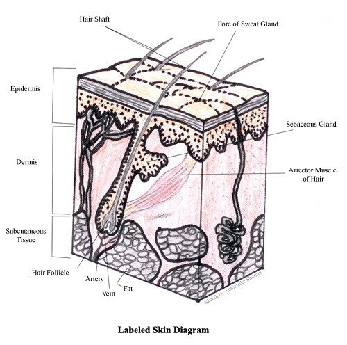
Doesn’t it just seem like every little piece of your body ends up being a thousand times more complex than you think? Well, that’s because this IS SCIENCE! And that’s just how we do things around here!
